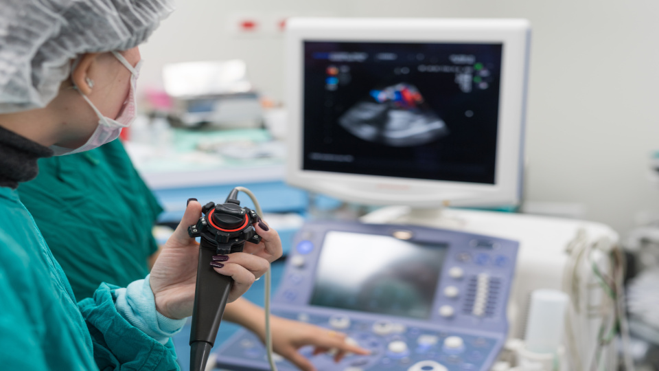
For the transesophageal echocardiography (TEE), a probe is inserted into the esophagus, similar to a gastroscopy, which allows very detailed ultrasound images to be taken of the heart, which is in direct contact with the esophagus.
This way the ultrasound probe is closer to the organ than when the examination is performed in the conventional way through the skin. In addition, the imaging is not affected by the lungs or ribs, which provides better resolution and therefore detail, especially in the area of the heart valves.
TEE makes it possible to detect or even rule out any blood clots in the heart, since this is the only method that can view areas of the atrium that are not visualized "from the outside" during an ultrasound examination. In addition, congenital heart defects can be precisely diagnosed.
As with a gastroscopy, patients receive brief sedation during the TEE, which takes about 15 minutes, and therefore should not drive themselves afterwards.
